Recognizing, Treating and Preventing Cracked Heels in Horses
Efrem Hunter, DVM
Efrem Hunter, DVM
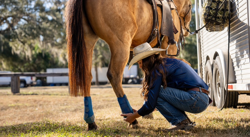
Cracked heels, also called scratches or mud fever, is a common name for pastern dermatitis, meaning inflammation of the skin on the back of the pastern (the part of a horse's leg between the fetlock joint and the hoof, which acts as a shock absorber) and fetlock (a crucial, high-motion joint on a horse's leg, often thought of as a horse’s “ankle”). As one of the more common lower leg issues in horses, early consultation with your veterinarian is recommended to help treat your horse and avoid future problems.
The term “cracked heels” is an informal name for pastern dermatitis. This condition is also known as scratches, greasy heel, mud fever, mud rash or dew poisoning. The inflammatory condition describes the location of the skin and appearance, not a specific cause. Bacterial infections, mites and blood vessel inflammation can all present similarly with lesions on the pastern, but each requires a different treatment approach. Identifying the underlying cause is essential, as similar lesions may need very different care.
Veterinarians refer to pastern dermatitis as a syndrome, indicating that it is a recurring pattern of lesions caused by several unrelated diseases. Lesions typically appear on the back of the pastern and fetlock, and may occasionally extend toward the cannon bone with one or multiple legs being affected. Cracked heels are often first noticed during grooming, when rough skin, redness or flaking is detected on the back of the leg instead of a smooth coat.
The underlying causes of lesions in this area can be grouped into several categories. Bacterial pastern infection, or pastern folliculitis, is most often caused by Staphylococcus bacteria, particularly Staphylococcus aureus, which has been identified in horses worldwide. Dermatophilus congolensis, the bacterium associated with rain rot, is another common cause, and fungal infections such as ringworm can produce similar lesions. When itching is prominent, Chorioptes mites may be responsible. These mites irritate the skin and cause itching, scaling and thickening, most often on the lower legs of draft-type and feathered breeds. Inflammation of the skin’s blood vessels (cutaneous vasculitis) is a separate cause.
Environmental factors, such as mud, wet pasture and damp bedding, can compromise the skin on the lower legs, so veterinarians routinely ask about housing, turnout and bedding conditions.
Cracked heels are easiest to address when the changes are still confined to the skin surface, and the back of the pastern is a spot most owners can check daily during grooming. The appearance of the lesions tells your veterinarian a great deal, because the same region is affected by chorioptic mange, dermatophytosis (ringworm), photosensitization (an abnormal skin reaction to sunlight), pastern leukocytoclastic vasculitis (blood vessel inflammation in the lower legs that sunlight often worsens) and pemphigus foliaceus (an immune-mediated skin disease). Those conditions, however, call for very different treatment.
The common signs of cracked heels include:
The first sign is often pink or red skin on the back of the pastern, frequently accompanied by small, raised bumps. This redness, known as erythema, may be followed by papules and pustules, which are small solid bumps and pus-filled lesions, a characteristic of bacterial pastern folliculitis. These early changes indicate that the lesions are still localized.
Dry flakes and thickened scabs commonly develop along the back of the pastern. Crusts, or dried material over inflamed skin, are often present. Circular areas of hair loss surrounded by scaling and crusting suggest an infection of the hair follicles, which may be fungal or a bacterial skin infection called pyoderma. Crusting that extends beyond the pasterns, including the coronary band (the vital line at the top of a horse's hoof where the haired skin of the lower leg transitions into the hard horn of the hoof wall), raises concern for pemphigus foliaceus, the most common autoimmune skin disease in horses.
Some horses develop moist areas where fluid seeps from the skin. Oozing is an early lesion seen in pastern leukocytoclastic vasculitis, often accompanied by erythema and crusting. When the discharge thickens and becomes purulent (suppuration), it signals that bacterial infection has advanced beyond the skin's surface. Oozing at this stage warrants prompt veterinary evaluation rather than ongoing observation.
Owners often notice splits or raw areas where the skin has broken open. An erosion is a partial loss of the skin surface, and an ulcer is a deeper defect that extends through it. In bacterial pastern folliculitis, individual pustules coalesce, meaning they merge into larger lesions, and untreated cases can spread into broad ulcerated areas that discharge pus. Erosions and ulcerations also appear in cutaneous vasculitis, often on unpigmented skin (white or pink skin without dark coloring).
Horses experiencing pruritus, or itchiness, may stamp, chew at the pastern or rub their legs against fences or posts. Itchy pastern dermatitis is often linked to Chorioptes mite infestation, especially in draft-type breeds, where these mites commonly target the lower legs. The severity of pruritus in cutaneous vasculitis varies and can be intense in some cases. When itching is the predominant symptom, particularly in draft breeds, mites become a leading diagnostic consideration for veterinarians.
Filling or puffiness in the affected leg is called edema, meaning fluid accumulation in the tissues. Edema in the leg arrives later in pastern leukocytoclastic vasculitis, after the crusting and ulceration stage. Swelling in more than one limb is also common in pemphigus foliaceus. Swelling appearing alongside crusting and ulceration is one of the more serious combinations and should prompt a call to your veterinarian.
Bacterial lesions turn sore once they reach the advanced stage. Movement is most likely to be affected after lesions progress to painful ulceration and suppuration, or once the whole lower leg becomes inflamed and swollen. Bacterial pastern infection rarely makes a horse sick in any other way, which leaves soreness in the leg as the only change many owners observe.
In long-standing cases, the skin of the pastern can become rough and develop a warty surface. This change is characteristic of chronic pastern leukocytoclastic vasculitis, a condition in mature horses that typically involves the pastern and hock regions, often affecting white or pink skin, though not exclusively. Lesions frequently appear on both sides of the pastern.

Diagnosis begins with a thorough history and physical examination. Your veterinarian will inquire about the horse’s age, breed, sex, living and working conditions, progression of the problem and all treatments previously attempted. After evaluating this information, your veterinarian will develop a list of potential causes and recommend further testing to confirm or eliminate specific conditions, which may include:
Skin scraping and grooming: Your veterinarian collects a surface sample and examines it under a microscope for mites, lice and fungal or yeast organisms. This test is often used when parasites are among the conditions being considered, which is common with itchy lower leg disease.
Cytology: Your veterinarian collects cells by impression smear, swab or fine needle aspirate and examines them to determine whether a lesion is infected, inflamed or related to a tumor.
Bacterial culture and susceptibility testing: A laboratory grows the organism from a sample to identify it and determine which drugs it responds to. Veterinarians recommend it for deep pyoderma and for surface pyoderma that has not responded to initial therapy.
Fungal culture or polymerase chain reaction (PCR) testing: These tests are used when lesions indicate dermatophytosis or when a fungal infection must be excluded due to atypical clinical signs. Fungal culture is a lengthy process, while PCR provides faster results, although its reliability can vary.
Biopsy: Your veterinarian removes a small skin sample for histopathology, meaning microscopic examination of tissue. Biopsy is often reserved for suspected immune-mediated disease, skin tumors and chronic or non-responsive cases.
Few skin diseases yield a definitive diagnosis based solely on diagnostic testing, so results must be interpreted in the context of the horse’s history and clinical findings.
Treatment should always be directed by a veterinarian, as cracked heels is a broad term that includes several distinct conditions, such as bacterial folliculitis, mite infestation, dermatophytosis, vasculitis and immune-mediated disease, with each requiring a specific therapy. Medications effective against one cause may be ineffective or even harmful for another. For example, corticosteroids are beneficial in immune-mediated disease but can worsen bacterial infections. Therefore, accurate diagnosis is essential before starting any treatment. Your veterinarian may remove hair from the affected area with clippers and wash the skin with a mild antiseptic before treatment begins.
For localized bacterial pastern lesions, topical therapy is applied directly to the affected skin. Topical treatments target existing lesions but do not address underlying causes that remain active on the skin. Your veterinarian will ensure that the selected topical therapy is appropriate for the specific diagnosis before starting treatment.
For instance, Equiderma Zinc Oxide Paste is a barrier paste for pastern dermatitis and lists chlorhexidine and miconazole, and it is appropriate after a veterinarian has identified the cause, since it does not address mites or immune-mediated disease. Another option, Banixx Wound and Hoof Care Horse and Pet Spray, lists scratches, mud fever and thrush among its uses.
Antiseptic shampoos that treat bacterial skin infections cover a broader area than ointments. Follow the label directions your veterinarian gives you for contact time and rinsing, and dry the leg thoroughly afterward. Shampoo therapy is a supportive measure within a plan, not a substitute for identifying the cause.
Systemic antibiotics are given by mouth or by injection to treat infection through the bloodstream rather than on the surface. They are warranted for deep pyoderma, for surface pyoderma that has not responded to initial therapy and for advanced lesions with ulceration and suppuration. Culture and susceptibility testing become especially important in these cases, because the results tell your veterinarian which drug the organism will respond to. Antibiotic selection is a veterinary decision and depends on the organism identified.
Antiparasitic medications are prescribed when Chorioptes mite infestation is diagnosed, particularly in draft-type breeds prone to itchy lower leg disease. These treatments eliminate the mites directly, addressing the root cause rather than merely reducing inflammation. As the infestation clears, skin condition typically improves. Your veterinarian will determine the most appropriate product and treatment schedule for each case.
Corticosteroids, administered orally or by injection, are the primary treatment for pastern leukocytoclastic vasculitis. In some cases, topical corticosteroid sprays can be used to reduce the required systemic dose. Pentoxifylline is another option that may be incorporated into the treatment plan. Management of pemphigus foliaceus also relies on corticosteroid therapy, sometimes supplemented with additional medications to minimize corticosteroid use.
Photo-aggravated vasculitis is worsened by ultraviolet (UV) light, and reducing UV exposure is part of treating it. Wrapping the legs blocks the light directly, and keeping the horse in a stall through the middle of the day reduces exposure (both are often recommended). This measure is only appropriate when photo-aggravation is confirmed, highlighting the importance of an accurate diagnosis before beginning treatment.

During recovery, the owner’s job is straightforward: follow the plan the veterinarian wrote, keep the environment clean and dry, watch the leg closely and report back on how it's responding. Resist the urge to guess at the cause yourself, switch products if progress feels slow or assume this case is the same as one you've dealt with before, since cracked heels can come from mites, bacteria or a blood vessel problem triggered by sunlight, and each needs its own treatment.
Prevention follows the same rule. There is no single product or vaccine that prevents cracked heels, because there is no single cause. A dry environment can help reduce moisture-related skin irritation and bacterial problems, but it does not help prevent the sun-triggered kind, which calls for shade or leg wraps instead.
Watch for lesions that are improving or worsening, a distribution that remains localized or spreads, itching and its severity and any restlessness, behavioral changes or reduced performance. Note any seasonal patterns, as that detail helps your veterinarian narrow down the cause. Report worsening lesions, spreading distribution, increasing pruritus or any response that falls short of what was expected. Leave crusts and scabs alone rather than picking or peeling at them, and give the prescribed medications time to work.
Give your horse a dry place to stand, away from mud and wet pasture, and keep bedding fresh and dry. A thick shavings bed, maintained with muck and stall cleaning tools, reduces irritation to the lower legs. If contact allergy is suspected, your veterinarian may suggest bedding that is not treated or aromatic. Turn your horse out once the grass has dried off, then clean and dry the lower legs at the end of the day.
Horse shampoos for skin and coat health, when recommended by your veterinarian, should be used according to the label directions. In feathered breeds, keeping the lower legs clean lets you see the skin and catch changes early, though evidence for clipping feathers as a preventive measure is limited, and that decision belongs to your veterinarian.
Some of the conditions that present as cracked heels are shared among horses, which makes barn management part of the plan. Chorioptic mange requires treating every horse in contact with the affected horse, not just the horse showing signs. With ringworm, the fungus travels on objects rather than through the air, which puts shared leg wraps, boots and grooming tools on the list. Ask your veterinarian which of these measures applies once the cause is known.
Frequently Asked Questions
No, and a horse that is current on routine vaccinations gains no added protection against it. There is no vaccine and no universal preventive product, which is not surprising given the number of causes grouped under the term. Prevention comes from environmental management and from addressing the specific cause in your horse.
No, this is not a reliable approach to treatment. Some cases are seasonal, some are worsened by sunlight, some are infectious, some are parasitic and some are immune-mediated. Drier conditions may help a case driven by the environment, but they will not resolve one driven by mites, bacteria or vasculitis. For the best outcome, work closely with your veterinarian to ensure your horse receives the appropriate diagnosis and treatment for its specific condition.
No. Cracked heels affect the skin of the pastern and heel, while a quarter crack is a split in the hoof wall that can start at the coronary band or at the ground surface and is managed by a veterinarian and farrier. The names sound related, but the conditions are not.
Author Summary Dr. Efrem Hunter Veterinarian Guest Author Dr. Efrem Hunter is a veterinarian, business owner, and pet industry consultant based in the Greater Houston, Texas, metropolitan area, where he owns and operates PetVet365-Cypress. He has more than 20 years of experience spanning clinical practice, pet nutrition, and the veterinary pharmaceutical industry. |